Tuesday, 18 December 2012

Neuroprosthetic controlled by thought


A woman, aged 52 and tetraplegic, has managed to control and manage a newly developed robotic hand with her thoughts only, U.S. scientists revealed in the journal The Lancet. Experts explained this advancement required a new form of transmission of nerve impulses that uses the natural control of muscles as a reference.

Jan had lost the ability to move her arms and legs more than 10 years ago due to a degenerative disease called spinocerebellar degeneration, which caused damage to his spinal cord similar to a fracture of the spinal column in a traffic accident. The team from the University of Pittsburgh, Pennsylvania (USA), directed by Andrew Schwartz, treated the patient before she was completely quadriplegic, although shortly after putting on the electrodes she could not longer move her arms. 

Scientists implanted her with two microelectrodes in the motor cortex of the brain. These were connected to a robotic arm with an artificial hand and fingers. Two days after the operation the woman could move the arm, from left to right, up and down, only through her thoughts. After 13 weeks of training, she could perform certain actions in order to take things and her movements became faster and more efficient, as scientists revealed.


The prosthetic arm, designed by the John Hopkins University's Applied Physics Laboratory. DARPA and JHU/APL​.

The innovative technique which makes use of the robotic arm is more intuitive for patients, because instead of having to think about where to move the arm, the patient simply has to focus on the goal, as one would do when trying to to score a basket. Although several groups around the world are developing similar prototypes, none has achieved these impressive results. Jan managed to move objects on a table, including cones, blocks and small balls, catching them and changing their location.

"We were impressed by how quickly she was able to acquire his skill. It was totally unexpected," said Andrew Schwartz, professor of neurobiology at the University of Pittsburgh and lead investigator of this project. "At the end of one day, when she was doing these beautiful movements, she was very excited."

To connect the woman to the robotic arm, doctors performed a four-hour operation to implant two tiny electrodes grids of 4 mm on either side of Jan's brain. Each grid has 96 tiny electrodes that protrude 1.5 mm . The electrodes were pushed just below the surface of the brain, near the neurons controlling the hand and arm movement in the motor cortex. Before Jan could use the arm, the doctors had to record her brain activity as she imagined various arm movements. For this, she was asked to look at the robotic arm and try to move it in the same way as she would do with her own arm.

What is yet to come

There are several challenges yet to solve. One is that the robot hand can send sensory impulses to people, so that they can interact with objects according to their texture and temperature.

A second objective is to develop thinner electrodes with a thickness of about five thousandths of a millimeter, to avoid problems like the one Jan has developed after the publication of the study. It is about a scar tissue that forms around the electrodes and that degrades the signals the brain sends to the computer. With smaller electrodes, the body may not trigger any healing process.

Another major focus of future work is to develop a wireless system, so that the patient does not have to be physically connected to the computer that controls the robot arm.

Wednesday, 28 November 2012

A robot controlled by thought


Researchers at the National Center for Scientific Research in France and the Japanese National Institute of Technology and Advanced Industrial Science at its Joint Robotics Laboratory (CNRS-AIST JRL) have been developing an EEG-controlled interface for a virtual avatar or physical robot. The goal is to train someday severely paralyzed people to operate a robot with their minds so it can bring things from home and maybe even clean the parts of the house.

The system is based on an artificial intelligence that understands the intentions of the user, so that the robot does not need to be micro-managed to perform simple tasks such as walking to the end of the corridor or lift a cup. Once the user focuses on a target, the robot knows what to do with it. 

Check it out in the following video in DigInfo: 

Friday, 23 November 2012

Visual implant helps the blind read Braille

A group of French and American researchers has succeeded in developing an ocular device that has allowed to transmit first braille patterns directly into the retina of a blind patient who has been able to read four letter words accurately and quickly.

"In this clinical test with a single blind patient, we bypassed the camera that is the usual input for the implant and directly stimulated the retina. Instead of feeling the braille on the tips of his fingers, the patient could see the patterns we projected and then read individual letters in less than a second with up to 89% accuracy," explains researcher Thomas Lauritzen, lead author of the paper.

The study, published in "Frontiers in Neuroscience, was conducted by researchers at Second Sight, the company that developed the device, called the Argus II, "the artificial retina", has already been mentioned in this blog in a post last April. As I said then, the concept is similar to the development of cochlear implants: there is a visual implant a grid of 60 electrodes attached to the retina to stimulate patterns directly on nerve cells. For this study, researchers stimulated six of these points on the grid to project braille letters.

argus II operation
Image: Copyright Gadget Review.

"There was no input except the electrode stimulation and the patient recognized the braille letters easily. This proves that the patient has good spatial resolution because he could easily distinguish between signals on different, individual electrodes." says Lauritzen.

The patient correctly identified 89 percent at one point, 80 percent in the case of two points, to 60 percent in the case of three words, and 70 percent of 4-letter words.

Source: 

Wednesday, 21 November 2012

More for the feet

As I am one of the many runners out there that use insoles to correct defective positions while walking and running, I found this quite interesting.

At the University of Utah engineering professor Stacy Bamberg recently developed a smart insole that seems to resemble the high technology Nike shoes and other Nike products for running. In this case, it goes beyond that, as this insole could help patients with prosthetic legs and people in rehab to correct walking abnormalities after a fracture or hip replacement.

The device is called Rapid Rehab and consists of a personalized gel insole embedded with force sensors, accelerometers and gyroscopes to detect the way a person walks. Rapid Rehab also contains a wireless transmitter to send data from sensors to a custom application for your smartphone that tracks and provides immediate visual, audio or sensory feedback to the user. The current version specifically contains two force-sensitive resistors that track the pressure the foot exerts when it is on the ground, as well as an accelerometer and a gyroscope to measure foot position and angle.

Image: copyright U News Utah

At present, the insole Rapid Rehab is used by amputees who want to reduce the limp when using a prosthetic leg. However, Bamberg expects it to be also used by physiotherapists and their patients who have received a hip replacement or suffered a bone fracture and need to correct their gait, since Rapid Rehab has proven to be less expensive than a gait-analysis laboratory study, more accurate than subjective observations of a physiotherapist, and faster in providing feedback.

Source: http://unews.utah.edu/news_releases/utah-engineering-prof-invents-smart-insole-to-correct-walking-abnormalities/

Wednesday, 14 November 2012

A bionic foot made in Brussels


I had not written for a long time about one of my favorite topics: bionic limbs. Fortunately this morning I found an interesting story that also touched me in a particular way, since the engineers who have developed this idea live in my city, Brussels.

Indeed, research engineers at the Vrije Universiteit Brussel, have developed a new active transtibial prosthesis that mimics the natural movement of the ankle and using energy efficiently. Instead of using powerful engines which start and stop at every step, the system activates an electric motor continuously to continuous stretch a rubber band, which in turn is used by the foot as a source of motive power.

By reducing the total energy requirement of the prosthesis, you can use smaller batteries, reducing the overall weight of the device. Moreover, the smaller engines, quieter and more efficient can also simplify the design and implementation.

Check out the video, it's amazing how well it resembles the motion of an actual foot!


Tuesday, 13 November 2012

Treating liver cancer

I would like to write today about two different approaches to treat liver cancer, which have been recently presented in the news, both coming from Southampton in UK.

I read about the first one last 12th of November in BBC News. It is about a "chemo-bath", consisting of isolating the organ so the chemotherapy drugs don't reach other parts of the body. In words of Dr Brian Stedman, a consultant interventional radiologist at Southampton General Hospital: "To cut off an organ from the body for 60 minutes, soak it in a high dose of drug and then filter the blood almost completely clean before returning is truly groundbreaking."

The procedure was baptised as Chemosaturation with Percutaneous Hepatic Perfusion (CS-PHP) and it was used for the first time in UK in two patients with a cancer that had spread to the liver. The method had already been used in Germany, Italy, Ireland, France and the US. Furthermore, a study carried out in the US showed that patients who received this treatment survived 5 times longer than patients who received the best alternative care. 


Image copyright: DELCATH SYSTEMS INC - FORM 8-K - EX-99.1 - EXHIBIT 99.1 - June 3, 2011

This approach could save lives of patients whose cancer has spread to the liver from a primary tumor, for example the melanoma of the eye. Once the cancer reaches the liver, there is no effective treatment and survival is usually no more than four months, with one in ten patients living for a year. However, with this new treatment the time these melanoma patients can live is extended without the disease progressing.

In this new treatment the chemo drug is infused directly into the liver via catheter into the artery. Blood in the veins leading out of the liver is then captured and filtered through a specially designed, double-balloon catheter to filter out the drug before the cleaned blood is returned to the body. The approach allows the drug to be delivered, at a higher dosage than usual, directly to the liver and target the cancer tumor there, but in a minimally invasive manner.

Secondly, researchers at the University of Southampton led by Professor Salim Khako hepatology and Aymen Al Shamkhani immunologist, reported on 12th October that they will investigate how to boost innate immunity in humans to treat liver cancer. The idea is to stimulate the participation of NK cells (Natural Killer cells) to eliminate hepatocellular carcinoma cells.

Professor Khako highlights that hepatocellular carcinoma accounts for 90% of all primary liver tumors and to date is a very difficult condition to treat, in addition to being one of the growing mortal diseases in the world.

In a first phase the professor proposed a clinical trial using these NK cells in the treatment. He announced that three types of patients will have the formation of this cell stimulated to find the most appropriate method. When the team manages to find the most efficient system to stimulate NK cells, trials will be carried out in order to determine the best treatment for liver cancer, the university reported.

The BBC article:

Friday, 9 November 2012

The power of your ear

For the first time, scientists have managed to connect a medical implant and feed it with the energy of a natural battery that we all carry in our inner ear. Yes, all mammals have in their inner ear an "endocochlear potential" similar to a battery. It is located in a chamber filled with ions that, with the aid of sound waves, produce electricity gradients which become auditory nerve signals processed by the brain. In words of the discovers at MIT: 

"The ear converts a mechanical force — the vibration of the eardrum — into an electrochemical signal that can be processed by the brain; the biological battery is the source of that signal’s current. Located in the part of the ear called the cochlea, the battery chamber is divided by a membrane, some of whose cells are specialized to pump ions. An imbalance of potassium and sodium ions on opposite sides of the membrane, together with the particular arrangement of the pumps, creates an electrical voltage." 

"We have known for 60 years that this battery exists and that it’s really important for normal hearing, but nobody has attempted to use this battery to power useful electronics." says Konstantina Stankovic, a researcher at Harvard-MIT Division of Health Sciencies and Technology. 

So far, they have managed to use this source of electricity to power a tiny device without affect hearing.


Image: Patrick P. Mercier - MIT website

This technique could be used to power small ear implants which assist people with hearing loss, to deliver drugs to the body or to feed other sensors. At present, the voltage and power are so weak that scientists had to design a mini converter circuit so the electricity could be exploited by a small electronic device (see the image for a close view into the device size).

Tuesday, 30 October 2012

To have your knees about you


Much has been said and written in the last months about the knee of the most famous tennis player in my country, Rafa Nadal. Thus, I would like to write today about recent advances in knee injuries diagnosis and knee replacement.

ConforMIS personalized knee replacement

ConforMIS is making knee replacement surgery more personal, creating custom knee implants that exactly match the patient's anatomy. The company uses a technology known as rapid prototyping plus additive manufacturing, which converts a three-dimensional computer design into a physical object. A similar tool that allows personalized implant design and creation is the Materialise Mimics Innovation Suite. ConforMIS believes that such implants can help perform the knee replacement surgery more quickly, more accurately and with less traumatic effects for the patient.

The knee replacement surgery repairs damage and relieve pain in patients with severe osteoarthritis or knee injuries. The total knee replacement involves the extraction of dead cartilage and bone from the surface of the knee joint, the hip bone, the shin bone and the patella, and then replace it with an artificial joint made of a combination of metal and plastic. A partial knee replacement can also be carried out in only one part of the joint.

Typically, the surgeon chooses an artificial joint among several options of different sizes. ConforMIS, however, creates a custom implant based on data provided by images of the knee joint of the patient, with a technology that the company callas iFit. ConforMIS transformed medical images coming from a CT or MRI scanner in a three-dimensional computer model with the aid of a computer design program (CAD), and then used the 3D model as a template to manufacture the implant.


Use infrared to determine knee injury

Applied measurement sensors based on infrared may be used to diagnose patients with injuries in the ligaments of the knee, said Ricardo Aguilar, Biomedical Engineering student at ITESM Campus Chihuahua.

The anterior cruciate ligament injury of the knee is a recurrent injury in athletes, as well as in automobile accidents, when the knee is impacted. The biggest problem however is not the difficulty of treating the injury, but the diagnosis of the degree of damage suffered by the patient, he said.

Currently the only way to diagnose the degree of injury is to perform surgery in the patient to detect low visual appreciation tissue conditions. The biomedical research center ITESM Campus Chihuahua started since April this year an investigation coordinated by the orthopedics at Hospital Christus Muguerza del Parque, to design a measurement system which allows detecting, through an analysis of "pivochips", the gravity of the existing knee injury.

In a study based on "pivochips", sensors of an electromyograph with accelerometers owned by the Biomedical Research Laboratory are connected to the patient's injured knee, which are capable of measuring muscle behavior and the affected area during some movements by using infrared sensors, so the degree of injury can be determined without the need to perform surgery.

Wednesday, 17 October 2012

OR Revolution

Brainlab a step ahead?

Brainlab has launched what we could describe as a super-sized iPad (42 inches) which allows manipulating medical images among other possibilities. They have called it "Buzz Digital OR", a system that integrates all intraoperative imaging. It can be used for viewing DICOM images, but it also performs video management, routing video between sources and destinations, and includes options for fast and easy documentation of surgical procedures. The HD display comes with an integrated sound system, webcam and microphone. It can connect with a multitude of video signals and route content to multiple, full-HD displays. All data can be relayed across the hospital IP network. Like the normal iPad, it has drag and drop functionality which, for example, makes easier to navigate through the different images. 

Indeed, 2012 is being a great year for Brainlab guys since last March they succeeded in winning the coveted 'red dot award', beating over 4515 designs in an international competition. For its benefits, Curve™ inspired the experts and was awarded the "red dot award: product design". Curve incorporates the latest in medical technology. This advanced surgical navigation system for the brain and the body includes the latest software for image-guided advanced 3D displays. Curve features a great ergonomics, two multi-touch terminals, digital HD, hi-fi and wi-fi.


Brainlab Digital Buzz, copyright Brainlab


Gesture Control Technology 

TedCas, a small Spanish company, has invented a system based on Kinect technology, the Xbox game console camera, which allows gestural control of computer applications in ORs and other environments, just as Tom Cruise did in the movie "Minority Report'. The application allows surgeons to use image guidance in the computer without touching it, thus decresing the possibility of bacterial transmission. The number of people who contract an infection each year in operating rooms is around 300,000 cases per year in Spain alone. If we talk about the European Union framework, this figure rises to four million, causing public administrations to spend billions. 

The Spanish system also helps to improve process efficiency in ORs or radiation therapy rooms. Right now there are three options: to have someone managing the computer from outside the room, or the surgeon comes out to do it himself, which is a bit tedious because you have to repeat the whole process of sterilization, or the screen is within the room itself which, despite being protected, remains a risk of infection. Thus, gestural control makes easier for the surgeon to control how the medical information is displayed during the operation. 

At Sunnybrook in Toronto surgeons can now benefit Kinect system during operations, as you can see in the video below:


Source: medgadget, http://www.brainlab.com/art/3401/6/brainlab-introduces-new-multi-touch-surgical-information-hub/

Monday, 1 October 2012

Medical device destroys itself leaving no trace in the body

Imagine an electronic device that releases the drug in the area where it is needed, measure your vital signs, treats a surgical wound and once done it has done its job it melts away ... All this without leaving a trace, as if it had never been in the body. A new class of devices grouped under the name of "transient electronics" is about to start a medical revolution. The first step was just taken by a group of engineers at Tufts University, in the United States, who successfully tested one of these biodegradable devices. The experiment was done with mice that were implanted with a device designed to deliver drugs and treat an infection. Not only did the treatment work, but also it barely left remmants of its presence in the organism three weeks later. With only a few tens of nanometers thick, the new circuits are easily dissolved in either water or other body fluids without causing damage.

In the same line, Abbot lab has recently introduced a new "stent", a new type of coronary implant which is also absorved when is no longer useful. It has a less sophisticated technology than that of the electronic devices, but it works similarily. Stents are a mesh-like tube of thin metal wire which are placed inside the artery to keep them open and facilitate blood flow after a heart attack has occurred. Stents have been placed permanently for years. The absorbable one, made ​​of a material used in sutures, allowes the vessel to dilate and contract in a more natural way, as your body needs it. For example when you are running and need more blood flow. At the end of ist lifetime, the stent is complety disolved and the blood vessel remains open without any other extra support.

Sources:


Monday, 17 September 2012

Understanding more the brain complexity


A brain implant could help regain the ability to make decisions, a process that may be lost after a stroke or brain injury, restore the neural mechanisms necessary for this process. According to a study published in the Journal of Neural Engineering, the placement of a neural device in the frontal lobe of a group of brain-injured monkeys succeeded in recovering, and even improving, their ability to make decisions.

Researchers at the University of Kentucky and the University of Southern California (USA) seem to have taken a key step towards the recovery of specific brain functions in patients with brain injuries. The study was based on a neural prosthesis implantation which evaluated the ability of neuronal communication in the brain of animals. 

Once that scientist obtained enough information about neuronal signaling and how this affected memory and recovery processes related to decision-making ability, they devised a model of multiple input, multiple output (MIMO) which stimulated those signaling neuronal pathways which had been and were necessary for the decision-making processes. The results showed that the MIMO model was highly effective in recovering the task performance capabilities and it was even able to improve performance in normal condition.


Wednesday, 5 September 2012

Walking again

A British woman paralyzed from the chest down after a riding accident has become the first person to use permanently a bionic exoskeleton to walk, a device that so far has only been used in hospitals and rehabilitation centers.

Claire Lomas is the first person who takes the suit 'ReWalk' home for a daily use, after having previously used it for 17 days to participate in the London Marathon, which he used to raise 200,000 pounds for backbone research. She has recognized that routine activities are very rewarding now as standing "means everything" to her. "One of the best experiences was standing in a bar," she said.

Larry Jasinki, CEO of Argo Medical Technologies, the company that developed the exoskeleton suit, explained to Reuters that at first he had his doubts about supporting participation in the race because the suit was still in test phase, but Lomas said she managed it well. "The costume was very reliable in the worst weather and, after 17 days, I arrived (to the target)," she said.

The person wearing the exoskeleton activates it by bending their body, indicating their desire to take a step. In addition to supporting the weight of the body, it allows the user to walk up or down stairs and sitting or standing independently.

The device costs around 45,000 pounds, and although ongoing clinical studies could support the idea that health authorities finance the purchase of the device, its developers indicate that savings in the treatment of health problems related to inactivity could offset the cost.

More information:


Monday, 7 May 2012

A mechanical hand controlled by thought

Reality overcomes fiction once more. Some years ago, a bionic arm was something we could only imagine in the Terminator or Star Wars movies. Remember the mechanical hand that Luke Skywalker had implanted after  Darth Vader cut his hand with his lightsaber? Well, nowadays thousands of people with amputees arms already benefit from a similar technology. 

Initially patients could move the arm through redirected nerves that sent electrical signals to sensors placed on his chest. The first prototype was implanted to the American Jesse Sullivan in 2002. Jesse is an electrician and suffered a terrible accident in May 2001 as he touched a wire with a voltage close to 7,500 volts. The bionic arm that he had implanted cost around six million dollars. The second person who received one of these prostheses was the young Claudia Mitchell (27 years). The former Marine lost his left arm in a motorcycle accident and has been operating with its robotic limb since August 2006. Claudia can now put on shoes, wash or peel a banana. Furthermore, she had the sense of touch restored as she was able to feel things she touched with her artificial hand through a device attached to her chest. During a four-hour operation, surgeons moved nerves from her shoulder, which normally ferry signals from the hand to the brain, and redirected them to muscles in her chest area. Four months after surgery, a patch of skin on her chest was able to feel touch, temperature and pain sensations as if they were coming from different parts of her hand and wrist.


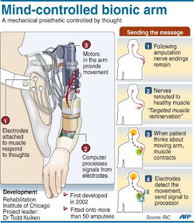
Last year, Dr. Todd Kuiken, director of bionic medicine center and director of amputees at the Rehabilitation Institute of Chicago (RIC) presented a new advancement, the Targeted Muscle Reinnervation (TMR). Kuiken and his team developed this innovative surgical procedure that routes signals from the brain to the nerves of the muscles that are healthy after an amputation, allowing patients to control their prosthesis just by thinking the action they want to perform. This was the world's first bionic arm controlled by nerves, which reflects the patient's brain impulses. (See image to learn a bit of how it works)


If you want to know more about bionic arms, I recommend you the Bionic Bodies series on the BBC News website: http://www.bbc.co.uk/news/health-17139965

Also, check out this video if you want to see how a young orthopaedic patient is getting used to a new bionic arm:


See you soon

Saturday, 7 April 2012

More on anticancer drug delivery using nanoparticles

I just came across an article in http://www.medicalnewstoday.com which goes over the use of  use of gold nanoparticles carrying drug molecules to target and treat cancer cells, something that was already presented in this post last February. This confirms that scientist keep foreseeing nanotechnology as new treatment against cancer, which might also replace radiation, chemotherapy, etc... in a near future.

In the study presented, the researchers used a single-stranded DNA aptamer called AS1411 which does two jobs. The first is to bind with the "shuttle" protein nucleolin, which is over-expressed in cancer cells and present inside the cell and on the cell surface. The second job is, once it is released from the nanostar, is to act as the drug itself. The results were quite more than satisfactory, as scientist observed death and decline in cancer cell population, exactly the intended objective.

You can find the whole article at this address: 
http://www.medicalnewstoday.com/articles/243856.php

See you in the next biomedipost :)

Tuesday, 3 April 2012

Never-before-seen implant

In my last post, I discussed about what we can expect from developments in cochlear implants, a technology that has been with us for more than 30 years. As it was briefly explained, cochlear implants stimulate the cochlea by means of electrodes, making possible for the brain to interpret this information as sound. In this way, I have been thinking if it wouldn't be possible to do the same for blind people. A quick search on google and... voila, I found that we already have what it is called "a retinal stimulator" or "artifical retina" implant.

Some history I found about it: In the mid-1980s, the neuroophthalmologist Joseph Rizzo III was researching retinal transplants to restore blind people's vision. One day, removing a lab animal's retina, a tissue-thin membrane that lines the back of the eyeball's interior, he made a tremendous discovery. "The moment I made the cut, I said to myself, 'What in the hell are you doing?'" Rizzo recounts. He realized he was cutting nerve connections that are actually spared in many forms of blindness. The retina's light-sensing cells (photoreceptors, or rods and cones) die off in retinitis pigmentosa and age-related macular degeneration, which affect millions worldwide; but the nearby neurons that ferry the signals from those cells to the brain remain intact.

Electrode implanted in the back of the eye (© Springer Science) 
So Rizzo conceived what he called "a retinal prosthesis", a device intended to bypass the damaged eye structure. This has evolved to what we call nowadays and artificial retine which works as follows: a miniature camera mounted in eyeglasses captures images and wirelessly sends the information to a microprocessor (worn on a belt) that converts the data to an electronic signal and transmits it to a receiver on the eye. The receiver sends the signals through a tiny, thin cable to the microelectrode array, stimulating it to emit pulses. The artificial retina device thus bypasses the damaged photoreceptor cells and transmits electrical signals directly to the retina’s remaining viable cells. The pulses travel to the optic nerve and, ultimately, to the brain, which perceives patterns of light and dark spots corresponding to the electrodes stimulated. Patients learn to interpret these visual patterns. A lot of research and effort has been put in this technology since then and finally last year the first such device was approved in Europe for commercialization: The Argus II Retinal Prosthesis System by Second Sight.

But this shouldn't be perceived as making possible for blind people to see. In the same way that cochlear implants only partially restore hearing, the artificial retinal is not intended to fully restore vision, but to artificially provide electrical signals that the brain can interpret as shapes. Research is made towards increasing the number of electrodes thus increasing the "reslution" of the images that can be perceived. The ultimate goal is to design a device with hundreds to more than a thousand microelectrodes (DOE Artificial Retina Project). This resolution will help restore limited vision that enables reading, unaided mobility, and facial recognition. Also, some research goes towards the use of human tissue to improve the communication between biological tissues and artificial sensors. Researchers in Italy have now reported the functional interfacing of an organic semiconductor with a network of cultured primary neurons. Their novel approach represents a new tool for neural active interfacing, which is a simpler alternative to the existing and widely used neuron optogenetic photostimulation techniques, and avoids gene transfer, which is potentially hazardous. In words of one of the researches, Guglielmo Lanzani, "This new approach to the optical stimulation of neurons may stimulate further work towards the development of an artificial retina based on organic materials."


Saturday, 17 March 2012

Cochlear implants... a successful history with a bright future ahead

A friend of mine said to me few days ago: cochlear implants are one of those things of medicine and technology which actually looks like "magic" or "a miracle". Indeed, cochlear implants made possible to connect deaf people with the world of sound, allowing them to converse with others and even to listen to music. A dream for deaf people which started to take shape for the first time in Dr. Graeme Clark's mind, an Australian otolaryngologist pioneer in the field. He was the first person to develop the multi-channel cochlear implant and to have successfully performed the world’s first implant procedure on Mr Rod Saunders in August 1978, at Melbourne’s Royal Victorian Eye and Ear Hospital.
 

But explaining the whole history of this hearing implants is not the purpose of today's post. I would ike just to mention some of the new technologies currently developped in this interesting field, just to show that there is still big room for improvement and that deaf people can hope for the best in upcoming years. Among others: 


- DACS: Direct Acoustical Cochlear Stimulation: implantable hearing device which bypasses the middle ear and applies powerful vibration directly at the oval window in order to stimulate the cochlea without significantly entering it. It replaces the function of the ear drum and ossicular chain with direct vibration at the oval window (take a look here for more information: http://www.acousticimplants.com/en/product/how-it-works.php)
- Increase number of electrodes: "hi fi" cochlear implant featuring 50 electrodes. It is hoped that the increased number of electrodes will enable users to perceive music and discern specific voices in noisy rooms.
- Laser stimulation: The laser stimulation produced more precise signals in that brain region than the electrical stimulation commonly used in cochlear implants. Laser stimulation is a promising technology for improving the auditory resolution of implants.
- Fully internal cochlear implant with an internal microphone system: a fully implanted system which can be worn all the time (sleeping, having a shower…). The ability to hear 24/7! As of April 2011, four people have undergone a trial of an internal microphone system, with two more yet to come. More information in this article: http://www.newscientist.com/article/mg21028064.800-ear-implants-for-the-deaf-with-no-strings-attached.html
- Liquid transmission means for transmitting acoustical energy to the cochlea, using a liquid filled tube which is surgically inserted through the middle ear to an orifice in the cochlea. The liquid filled tube acts as a transmission line with little losses because the liquid can be similar in acoustic properties to the perilymph in the cochlea and the impedance of the transmission line can be more closely matched to the acoustic impedance of the cochlea at the termination of the tube at the cochlea.
- Improvement speech processing strategies: In the n-of-m sound coding strategy, the signal is processed through m bandpass filters from which only the n maximum envelope amplitudes are selected for stimulation (m>n). While this maximum selection criterion, adopted in the advanced combination encoder strategy, works well in quiet, it can be problematic in noise as it is sensitive to the spectral composition of the input signal and does not account for situations in which the masker completely dominates the target. A new selection criterion could be based on the signal-to-noise ratio (SNR) of individual channels.
- Possibility of delivering drugs to the cochlea by means of the cochlear implant to develop a range of local pharmacologic interventions to prevent hearing degeneration.

I hope this is enough to arouse curiosity... see you in the next biomedipost.


If you want to know how the natural process of hearing works, click here... And also a nice video about cochlear implants from Cochlear Limited:

Sunday, 4 March 2012

The engine of life

As hard as it is to find nowadays anything that can last more than just a few years, our heart is there to prove that nature is capable of creating the most amazing and reliable things. The muscle that never rests, pumping and beating to keep every part of our body oxygenated, starting as soon as 22 days after we are conceived, just until the day we leave this world.  It beats more than 30 millon times per year and more than 2000 million times during a whole life in average. With each heart beat, it pumps 80ml of blood, making 8000l a day, the power enough to push a car over 32kms. And all this in a muscle with the size of a fist.

CT Scan of a human heart
Heart keeps representing a challenge to medicine, as cardiovascular disease is still the leading cause of death in the world, even above cancer or traffic accidents. An estimated 17 million people die of CVDs, particularly heart attacks and strokes, every year. Many of this deaths are related to blocked arteries problems, and today I would like to present two promising technological advances to fight heart disease.

The first is a new application in transmyocardial revascularization (TMR) laser technology that may offer an alternative method of treatment to patients with severe heart disease who are not candidates for coronary bypass surgery or balloon angioplasty. A normal heart depends primarily on the coronary arteries to deliver its blood supply from the left ventricle cavity, the pumping chamber of the heart. In patients with heart disease, the coronary arteries are blocked preventing normal blood flow to the heart muscle. However, they still have a large supply of oxygenated blood in their left ventricular cavity. For a subset of patients who are not candidates for traditional cardiac surgery, which bypasses blocked arteries, surgeons have to create new pathways for the blood flow. TMR uses laser energy to create these pathways through a series of 1mm channels from the outer surface of the heart through the heart muscle into the left ventricular cavity, allowing for an increased blood flow directly from this "blood-filled" chamber to the oxygen-starved areas of the heart muscle.

The second is related to something that has already been discussed in the blog: nanotechnology. Concretely, targeted nanoparticles callled nanoburrs that can reach damaged vascular tissue and may be used to deliver drugs that help clear arteries. This new particles have been designed by MIT and Harvard researchers with the aim of treating cardiovascular disease. The nanoburrs are coated with tiny protein fragments that allow them to stick to damaged arterial walls. Once stuck, they can release drugs such paclitaxel, which inhibits cell division and helps prevent growth of scar tissue that can clog arteries. Moreover, drugs are delivered over a longer period of time, and can be injected intravenously, preventing patients from enduring repeated and surgically invasive injections directly into the area that requires treatment.

In the next posts we will see what is going on with pacemakers, apart from other curiosities about the heart. In the meantime, take well care of your heart by doing regular exercise, cutting down on cigarettes and eating healthier. Surely it will make the difference.

Thursday, 23 February 2012

A pharmacy inside the body

The results of tests conducted by researchers at the Massachusetts Institute of Technology (MIT) and the company MicroCHIPS in a group of women with osteoporosis opens the door to the widespread use of remote administration of drugs. This was achieved by inserting a microchip under the skin which is capable of administering a medicine, thus allowing the patient to avoid daily injections of drugs.


And can’t patches do that already? Well, there are two main differences: the microchip allows remote control of drug release and also it can be used to administer more than one drug. The chip acts like a pharmacy inside the human body, a technology that can be used to treat from cancer to multiple sclerosis. Thinking further, I also envisaged the possibility of using the chip to administer vaccines automatically at the different stages of human development. And all this in a device with the size of a small pen drive!

And how does it work? The chip contains a daily dose of medication in small wells that are covered by a thin layer of gold nanoparticles which protects and prevents the drug from coming out.  The chip can be programmed to administer the medication according to a programmed schedule or commands sent wirelessly through a special frequency. These commands cause the gold to dissolve and allow the drug entering the bloodstream.

For testing, a group of seven women aged between 65 and 70 had the chip implanted through a simple procedure performed under local anesthesia. For four months the chip administered them an osteoporosis drug called teriparatide in doses comparable to those provided by injections, without any negative side effects.

“We hope this really is the dawn of a whole new way of thinking about delivering medications,” said co-author Robert Langer, a professor of cancer research at the Massachusetts Institute of Technology.  However, for he and his and colleagues, “the ‘hairpin’ road to the clinic might be long and winding, but a versatile implantable device that exploits the microchip approach for controlled drug delivery will be well worth the wait for patients with chronic diseases,”

Sunday, 12 February 2012

How doctors choose to die - by Ken Murray

Some food for thought... A provoking and touching article that was published in the guardian this week. The article approaches the controversial topic of dying with dignity, giving up to death in light of a decent quality of life in your final days in earth, instead of trying every possible treatment in a last attempt of surviving. Also, it made me think once more that my time in this world is limited, something that my current healthy state makes me forget.


http://gu.com/p/35bcj


How doctors choose to die

When faced with a terminal illness, medical professionals, who know the limits of modern medicine, often opt out of life-prolonging treatment. An American doctor explains why the best death can be the least medicated – and the art of dying peacefully, at home


A doctor
'Doctors know enough about death to know what all people fear most: dying in pain, and dying alone.' Photograph: Microzoa/Getty Images
Years ago, Charlie, a highly respected orthopaedist and a mentor of mine, found a lump in his stomach. He asked a surgeon to explore the area, and the diagnosis was pancreatic cancer. This surgeon was one of the best in the country. He had even invented a new procedure for this exact cancer that could triple a patient's five-year-survival odds – from five per cent to 15% – albeit with a poor quality of life. Charlie was uninterested. He went home the next day, closed his practice, and never set foot in a hospital again. He focused on spending time with his family and feeling as good as possible. Several months later, he died at home. He received no chemotherapy, radiation, or surgical treatment. Medicare didn't spend much on him.
It's not a frequent topic of discussion, but doctors die, too. And they don't die like the rest of us. What's unusual about them is not how much treatment they get compared to most Americans, but how little. For all the time they spend fending off the deaths of others, they tend to be fairly serene when faced with death themselves. They know exactly what is going to happen, they know the choices, and they generally have access to any sort of medical care they could want. But they go gently.
Of course, doctors don't want to die; they want to live. But they know enough about modern medicine to know its limits. And they know enough about death to know what all people fear most: dying in pain, and dying alone. They've talked about this with their families. They want to be sure, when the time comes, that no heroic measures will happen – that they will never experience, during their last moments on earth, someone breaking their ribs in an attempt to resuscitate them with CPR (that's what happens if CPR is done right).
Almost all medical professionals have seen what we call "futile care" being performed on people. That's when doctors bring the cutting edge of technology to bear on a grievously ill person near the end of life. The patient will be cut open, perforated with tubes, hooked up to machines, and assaulted with drugs. All of this occurs in the intensive care unit at a cost of tens of thousands of dollars a day. What it buys is misery we would not inflict on a terrorist. I cannot count the number of times fellow physicians have told me, in words that vary only slightly: "Promise me that if you find me like this you'll kill me." They mean it. Some medical personnel wear medallions stamped "NO CODE" to tell physicians not to perform CPR on them. I have even seen it as a tattoo.
To administer medical care that makes people suffer is anguishing. Physicians are trained to gather information without revealing any of their own feelings, but in private, among fellow doctors, they'll vent. "How can anyone do that to their family members?" they'll ask. I suspect it's one reason physicians have higher rates of alcohol abuse and depression than professionals in most other fields. I know it's one reason I stopped participating in hospital care for the last 10 years of my practice.
How has it come to this – that doctors administer so much care that they wouldn't want for themselves? The simple, or not-so-simple, answer is this: patients, doctors, and the system.
To see how patients play a role, imagine a scenario in which someone has lost consciousness and been admitted to hospital. As is so often the case, no one has made a plan for this situation, and shocked and scared family members find themselves caught up in a maze of choices. They're overwhelmed. When doctors ask if they want "everything" done, they answer yes. Then the nightmare begins. Sometimes, a family really means "do everything," but often they just mean "do everything that's reasonable". For their part, doctors told to do "everything" will do it, whether it is reasonable or not.
That scenario is a common one. Feeding into the problem are unrealistic expectations of what doctors can accomplish. Many people think of CPR as a reliable lifesaver when, in fact, the results are usually poor. I've had hundreds of people brought to me after getting CPR. Exactly one, a healthy man who'd had no heart troubles (for those who want specifics, he had a "tension pneumothorax"), walked out of the hospital. If a patient suffers from severe illness, old age, or a terminal disease, the odds of a good outcome from CPR are infinitesimal, while the odds of suffering are overwhelming. But, of course, doctors play an enabling role here, too. The trouble is that even doctors who hate to administer futile care must find a way to address the wishes of patients and families. Imagine, once again, the A&E ward with those grieving, possibly hysterical, family members. They do not know the doctor. Establishing trust and confidence under such circumstances is a very delicate thing. People are prepared to think the doctor is acting out of base motives, trying to save time, or money, or effort, especially if the doctor is advising against further treatment.
Some doctors are stronger communicators than others, and some doctors are more adamant, but the pressures they all face are similar. When I faced circumstances involving end-of-life choices, I adopted the approach of laying out only the options that I thought were reasonable (as I would in any situation) as early in the process as possible. When patients or families brought up unreasonable choices, I would discuss the issue in layman's terms that portrayed the downsides clearly. If patients or families still insisted on treatments I considered pointless or harmful, I would offer to transfer their care to another doctor or hospital.
Should I have been more forceful at times? I know that some of those transfers still haunt me. One of the patients of whom I was most fond was a lawyer from a famous political family. She had severe diabetes and terrible circulation, and, at one point, she developed a painful sore on her foot. Knowing the hazards of hospitals, I did everything I could to keep her from resorting to surgery. Still, she sought out outside experts with whom I had no relationship. Not knowing as much about her as I did, they decided to perform bypass surgery on her chronically clogged blood vessels in both legs. This didn't restore her circulation, and the surgical wounds wouldn't heal. Her feet became gangrenous, and she endured bilateral leg amputations. Two weeks later, in the famous medical centre in which all this had occurred, she died.
It's easy to find fault with both doctors and patients in such stories, but in many ways all the parties are victims of a larger system that encourages excessive treatment. Many doctors are fearful of litigation and do whatever they're asked to avoid getting in trouble. Even when the right preparations have been made, the system can still swallow people up. One of my patients was a man named Jack, a 78-year-old who had been ill for years and undergone about 15 major surgical procedures. He explained to me that he never, under any circumstances, wanted to be placed on life support machines again. One Saturday, however, Jack suffered a massive stroke and was admitted to A&E unconscious, without his wife. Doctors did everything possible to resuscitate him and put him on life support. This was Jack's worst nightmare. When I arrived at the hospital and took over Jack's care, I spoke to his wife and to hospital staff, bringing in my office notes with his care preferences. Then I turned off the life support machines and sat with him. He died two hours later.
Even with all his wishes documented, Jack hadn't died as he'd hoped. The system had intervened. One of the nurses, I later found out, even reported my unplugging of Jack to the authorities as a possible homicide. Nothing came of it, of course; Jack's wishes had been spelled out explicitly, and he'd left the paperwork to prove it. But the prospect of a police investigation is terrifying for any physician. I could far more easily have left Jack on life support against his stated wishes, prolonging his life, and his suffering, a few more weeks. I would even have made a little more money, and Medicare would have ended up with an additional $500,000 (£314,500) bill. It's no wonder many doctors err on the side of over-treatment.
But doctors still don't over-treat themselves. Almost anyone can find a way to die in peace at home, and pain can be managed better than ever. Hospice care, which focuses on providing terminally ill patients with comfort and dignity rather than on futile cures, provides most people with much better final days. Amazingly, studies have found that people placed in hospice care often live longer than people with the same disease who are seeking active cures.
Several years ago, my older cousin Torch (born at home by the light of a flashlight) had a seizure that turned out to be the result of lung cancer that had gone to his brain. I arranged for him to see various specialists, and we learned that with aggressive treatment of his condition, including three to five hospital visits a week for chemotherapy, he would live perhaps four months. Ultimately, Torch decided against any treatment and simply took pills for brain swelling. He moved in with me.
We spent the next eight months having fun together like we hadn't had in decades. We went to Disneyland, his first time. We'd hang out at home. Torch was a sport nut, and he was very happy to watch sport and eat my cooking. He even gained a bit of weight, eating his favourite foods rather than hospital food. He had no serious pain, and he remained high-spirited. One day, he didn't wake up. He spent the next three days in a coma-like sleep and then died. The cost of his medical care for those eight months, for the one drug he was taking, was about $20.
Torch was no doctor, but he knew he wanted a life of quality, not just quantity. Don't most of us? If there is a state-of-the-art of end-of-life care, it is this: death with dignity. As for me, my physician has my choices. There will be no heroics, and I will go gentle into that good night.
• Ken Murray, MD, is clinical assistant professor of family medicine at USC. Taken from an article originally published at Zócalo Public Square.

Saturday, 11 February 2012

Mobile Health, healthcare for everyone, everywhere

Mobile technology is one of the fastest growing telecommunication industry of the world. It is a fact that, even in poor countries, this technology has experienced tremendous growth, as it is estimated that their population owns more than 2.200 million mobiles. However, the access to a better medical service is still limited, since the developing countries still lack medical professionals and beds in the hospitals. This is how the mHealth was born, referring to the “application of mobile communications and network technologies for healthcare“. mHealth allows to make the most of the current mobile technology to improve healthcare worldwide, especially in emerging countries.

As a result of the emergence of the mHealth field, a first alliance was created in order to address the challenges raised by this new vision of Healthcare. Thus, the mHealth Alliance was established with the purpose of supporting further research and development in the area. The founding fathers committed to create a non-profit mHealth coalition to maximize the impact of mobile technologies on health, especially in emerging economies. The Alliance is hosted by the United Nations Foundation, and was founded by the Rockefeller Foundation, Vodafone Foundation, and UN Foundation. As a result of this Alliance many projects have been started, being the so-called “mHealth for development” one of the most important. This project involves the United Nations Foundation together with the Vodafone Foundation and it is intended to “support the use of rapid response mobile telecommunications to aid disaster relief; to develop health data systems that improve access to health data thereby helping to combat disease; and to promote research and innovative initiatives using technology as an agent and tool for international development”. 26 countries have benefited so far from this initiative, including India, Uganda and South Africa, just to mention a few.


Other interesting projects and mobile health applications include: DataDayne (use of PDAs and mobiles to collect data such as nutritional statistics, medical supply change, immunization and vaccination programs in the field), Text2Change as well as Project Masilulekeis (to spread awareness and education about particular communicative diseases amongst large populations by sending SMS) and SIMPill (a pillbox which is fitted with a SIM card so that a healthcare worker is alerted when the pillbox is opened, letting him know that the patient is taking the medicine). 


And what’s there for the future? Check reports and videos  from the 3rd mHealth Summit which took place in December 2011. The idea was to organise different conferences to explore, examine, and debate the ways mobile technology will transform health care delivery, research, business and policy for the 21st Century. More information and highlights:

Sources: