Wednesday 18 December 2013

Ophthalmologic adapter for iPhone

Welch Allyn received clearance from FDA for its application iExaminer, which connects the iPhone to a PanOptic ophthalmoscope, providing more possibilities and useful information on examination of the fundus. This combined system allows doctors to take pictures of the eye and also save images for later review or to share with colleagues. Furthermore, the system is essentially a telemedicine product that can make possible for ophthalmologists to reach remote clinics missing an eye care specialist.

The iExaminer provides an optical alignment with the eyepiece of the ophthalmoscope and the camera of the phone and is compatible with the iPhone 4 and 4S models. There is also a complementary application of Welch Allyn to complete the package, and is available for download in the Apple App Store.

More info in the website of the product:

Copyright Welch Allyn


Tuesday 10 December 2013

Mobile DNA analysis in real time through your smartphone

More on mobile health technology.

Biomeme, a new company from Philadelphia, is planning to launch an easy to use system that can perform a real-time polymerase chain reaction (qPCR is its acronym in English ) and provide results through any smartphone, without using expensive lab equipment or an expert to work in it. The goal is to provide medical professionals, regardless of their location, the ability to perform advanced diagnostics and monitoring diseases.

Biomeme platform transforms a smartphone into a mobile laboratory for advanced DNA diagnostic and disease monitoring in real time. The system includes:

- A docking station for the real-time polymerase chain reaction (qPCR)

- A mobile application to control the system through wireless connection, analyze the results in real time and send them to the cloud

- Target test equipment for sample preparation and identification of pathogens or diseases, according to its specific DNA or distinctive RNA.

This low cost system requires no special laboratory equipment and could allow mobile inspection at a healthcare point, agriculture, monitoring of vectors, veterinary medicine, environmental monitoring, and even education.

More info here: http://bio-meme.com/

Biomeme Device Picture
Copyright bio-meme

Tuesday 3 December 2013

Building a smartscope

Smartphones are becoming more and more important in the medical sector, ranging from mHealth to medical applications which can turn your smartphone into a medical device. In this post I would like to talk about what someone has recently achieved by just using a Smartphone and some creativity: user Yoshinok from Instructables web site has managed to turn a smartphone into a microscope.

Well technically he didn't use just a smartphone. You'll need also a small box, a LED light and a laser pointer lens. With all this in place, you can increase the zoom of your smartphone 175x, allowing you to observe plant cells and details of objects, like a real microscope.

The invention seems to work perfectly. At least, in the video the user gets to see cells and details of coins, among other things. It also allows you to take a photo or video of the analyzed object... This invention only proves that when you use your creativity the possibilities are endless.


Wednesday 16 October 2013

Mobile Diagnosis via Smartphone

While many companies are using mobile analytics to measure their mobile advertising and marketing campaigns, scientists are developing healthcare-related smartphone apps that could actually save lives. The new apps are designed to help doctors by integrating with medical devices, enabling the smartphone to become the tool for data handling, analytics, visualization and communication.

For example, scientists at the Center for Systems Biology at Massachusetts General Hospital have integrating a microNMR (nuclear magnetic resonance) device that accurately detects cancer cells with a smartphone, according to the article.

Another example was recently presented at Biotechnia 2013 by the Fraunhofer Institute for Applied Information Technology (FIT): the nanopotentiostat, an Ambient Assisted Living (AAL) system for monitoring age-related risk factors. AAL systems continuously monitor patients at home and are networked with medical centers to enable medical observation and diagnosis without inconvenient visits to the doctor. In the case of the nanopotentiostat, one of its major features is that it integrates three different sensors in one platform.

This mini all-rounder can determine the blood sugar, lactic acid or cholesterol from a single patient sample, use fluorescence to detect selected biomarkers and record the patient’s heart frequency and oxygen saturation. The data is processed via an app and forwarded securely to the patient’s doctor. This system has been developed by Fraunhofer FIT in cooperation with Charité and T-Systems Deutschland in the MAS (Nanoelectronics for Mobile AAL Systems) project that is sponsored by the EU and the German Federal Ministry of Education and Research.

Further information:

Wednesday 24 July 2013

Biomaterials of the future


A group of scientists from the Department of Chemical Engineering, University of Michigan (USA) has discovered a new material made of spherical gold nanoparticles which could become the best flexible electricity conductor ever designed to date. "Essentially the new materials of metal nanoparticles behave as elastic. This is just the beginning of a new family of materials that can be manufactured from a variety of nanoparticles for a wide range of applications," explains lead author, Nicholas Kotov.

The elastic conducting materials research today is driven by technology needs. For example, we would all like to have flexible mobile phones or tablets that do not break if dropped. But some of the most important applications envisioned for these new materials in the future have more to do with medical implants that can move with the body.

Since graphene was first synthesized, something which earned Andre Geim and Konstantin Novoselov the Nobel Prize in Physics in 2010, it seems that this material built from carbon will solve all the challenges that scientists face nowadays. But, though their properties are amazing, definitely it is not the only flexible conductor currently being investigated.


According to the authors of the publication that has just appeared in the journal 'Nature', the possibilities range from cardiac implants capable of transmitting electrical impulses of the heart and move with the cardiac muscle itself to brain electrodes or flexible electronics. As cited in the work, the conductivities of electricity achieved with this material made of five layers of gold are similar to those of mercury, which is a great conductor. This property makes Kotov and his colleagues see this material as a candidate for the manufacture of electrodes, in particular, Kotov is interested in further research on the development of brain implants. "These can alleviate many diseases, such as severe depression, Alzheimer's or Parkinson's. It can also serve as part of artificial limbs and other prostheses controlled by the brain," says Kotov.

In the same vein, researchers at the University of Berkeley have uncovered one of the most complex electronic systems ever built on plastic. The invention consists of a thin plastic sheet that emits light with an intensity that reflects just the right amount of pressure applied to its surface, giving clues about how interfaces for flexible computers could be designed in the future. Described in the journal Nature Materials, the new light emitting electronic "skin" (that's how it is called by its inventors), is an extension of previous work Ali Javey lab, professor of electrical and computer engineering at the University of California at Berkeley. Javey group has developed a process which uses a large number of traditional manufacturing techniques for silicon uniformly and reliably integrating various organic and inorganic constituents on a plastic.

Thursday 20 June 2013

Human brain atlas with highest resolution

The dream of a neuroscientist is to have an accurare representation of the human brain and now we are closer than ever to that ideal with BigBrain, a digital reconstruction of the human brain full 3D and ultra-high resolution. BigBrain is the essential tool neurological laboratories worldwide need in order to elucidate the form and function of our brains.

Processing the brain layers - Amunts, Zilles, Evan et al (Science)

It is true that there are currently other brain atlas, but they only arrive at the macroscopic level, or visible. Its resolution only reaches the level of a cubic millimeter, and in that volume of brain, 1,000 neurons can easily fit. The new BigBrain offers a resolution very close to cellular dimension, according to scientists who have created it. That means you get to discriminate each small circuit of neurons that is behind our mental activity, which may include all available information on the brain, from genes and neurotransmitter receptors to cognition and behavior.

To construct BigBrain, researchers have taken samples from a patient 7400. The reference brain is based on a woman who died at age 65, which was sliced in 7,400 histological sections of just 20 microns. The BigBrain, according to its creators, opens the way for understanding the neurobiological basis of cognition, language and emotion, and also to investigate neurological diseases and develop drugs against them. The model is presented in Science and will be available for registered users at http://bigbrain.cbrain.mcgill.ca.

Tuesday 11 June 2013

First implant of a bioartificial blood vessel

Doctors at Duke University Hospital have announced the first successful implant of a bioartificial blood vessel created in the laboratory by the company Humacyte. The first patient to receive the implant was a man of 62 years with kidney failure who, after two hours of surgery, received a vein graft on 5 June.
The vein was designed using donated human cells growing on a scaffold tube to form a container that is specially treated to clean all the qualities that might trigger an immune response rejection.

"In pre-clinical testing, the veins have performed better than other synthetic implants and those of animal origin. This is a pioneering event in medicine, "said Jeffrey H. Lawson, MD, PhD, a vascular surgeon and vascular biologist at Duke University. The Food and Drug Administration (FDA) recently approved a phase 1 trial involving 20 patients on dialysis in the United States. Initial testing focuses on the implantation of blood vessels in a convenient place in the arms of renal patients on hemodialysis. 

vena

Initially, the researchers sought to develop veins using a person's own cells growing in a scaffold, reducing the risk of implanted tissue rejection in the patient's body. But because of the long time required for mass production this method was discarded, so that investigators changed course to develop a universal product using donated human tissue.

Million people worldwide require hemodialysis which often needs a graft to connect an artery to a vein to accelerate the flow of blood during treatment. Current options have drawbacks as synthetic vascular grafts are prone to clotting, causing frequent hospitalizations. This graft also could be used for coronary bypass or to replace blocked vessels in limbs. With no doubt it is a great advance in medicine that promises many benefits.

Tuesday 28 May 2013

Micro robots in the eye

One of the leading causes of blindness in many people is oxygen deficiency in the retina, and what is even worse, ophthalmologists do not have proper tools to make the correct diagnosis, which is extremely important when it comes to an organ as delicate as an eye. But what if all that trouble could be spared by tiny robots which were able to detect the deficiency of oxygen in the retina? Precisely that is what scientists at ETH Zurich are developing, specifically Prof. Bradley Nelson, director of the Institute of Robotics and Intelligent Systems, who also has a Guinness record for the most advanced mini robot for medical use.

The professor says that he sees these robots as similar to those used today to explore the seabed, being the major difference the fact they can measure levels of oxygen in the fluid of the eye and retina. They operate based on the behavior of a fluorescent dye which reacts to the amount of oxygen in the surroundings and, when stimulated with certain wavelengths of light, the dye emits fluorescent light which begins to disappear at a certain rate depending on the amount of oxygen present that is, the more oxygen the faster light disappears.

At the moment the tests were performed in water and the eyes of dead animals, but the results obtained are believed to make them ready for use in the human eye. Thus we might see in the near future ophthalmologist making use of robots to diagnose patients and find the best ways to prevent blindness.

http://www.engineering.com/DesignerEdge/DesignerEdgeArticles/ArticleID/5700/Micro-Robots-Could-Prevent-Blindness.aspx?goback=%2Egde_1817500_member_239595102


Thursday 23 May 2013

Bioresorbable Airway Splint


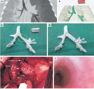
A two-month-old baby has saved his life upon being implanted with a splint in the trachea in a hospital in Michigan (USA), which was reproduced in a 3D printer, as published today in The New England Journal of Medicine.

The newborn suffered constant attacks caused by a syndrom called tracheobronchomalacia, which prevents oxygen from reaching the lungs due to a problem in the trachea problem, as diagnosed by doctors who have published the study. In view of the situation, doctors decided to print a kind of splint, which reproduced the tracheal tube based on a tomographic image of the child's airway.

The 3D reproduction is made with a material called polycaprolactone, bioabsorbable by the human body in three years, so the patient doesn't need another surgery to have the implant removed. The implant was reproduced on the printer in less than a day.

According to doctors, when the implant is removed naturally, the child's lungs and airways will be developed enough to stay open by themselves. After inserting the tube, doctors kept the baby on life support for 21 days, after which the baby was allowed to leave the hospital. One year after implantation, there have been no rejection or any problem with breathing.

This case demonstrates, doctors conclude in the study, that the combination of high resolution images plus the computer design and biomaterials for 3D printing can facilitate the creation of precise anatomical implants. The study has been signed by Scott Hollister and Richard Ohye from the University of Michigan, and Marc Nelson from Akron Children's Hospital.


The three-dimensional printers are making headway in medicine, but for now the applications are very limited. Initially, they were used to design conventional solid materials intended to design personalised implant prosthesis. One of the benefits is the ability to create custom models at lower prices than conventional industrial processes. The next step, as the case published today in the New England Journal of Medicine, is to design objects with a biological material such as the biodegradable polymer employed in the intervention to reproduce the trachea fragment that the patient was missing.

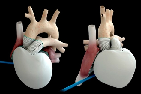
The ability to create these structures combined with the manipulation of stem cells could lead in the future to the creation of artificial organs. At least, scientist are working in this direction. A biological mold would be the basis to provide a three-dimensional structure to the organ. The outer casing would be scattered on cells which would colonize the structure resulting in specialized cells of the organ concerned (cardiomyocytes in the case of the heart, hepatomiocitos for the liver...).

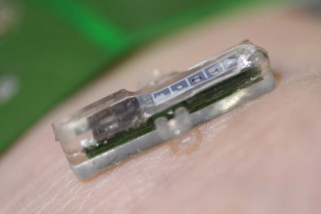
This possibility remains part of science fiction. What is beginning to be a reality is a new generation of 3D printers that generates human tissue. The company Organovo announced last April at the Experimental Biology Conference in Boston an equipment capable of producing tiny liver tissue samples (about 20 cell layers thick). This printer uses biological ink which in this case is made of three types of cells: hepatocytes, stellate cells and cells from the walls of blood vessels, obtained from waste coming from transplants and surgical interventions. Currently, the main use of these miniature livers is to test the effectiveness and impact of new drugs, but it may be the first step in a future technology able to print of organs for transplantation.

http://www.nejm.org/doi/full/10.1056/NEJMc1206319

Tuesday 14 May 2013

The most advanced bioartificial heart


The French company Carmat has been authorized to begin implementing its new human artificial heart, considered one of the most advanced to date. Although only some pilot tests have been carried out so far, the new artificial organ will be used for the first time in four hospitals in Belgium, Poland, Slovenia and Saudi Arabia.

This model is considered the latest generation of artificial heart, and although there are many advantages that have been observed in the tests, it is also true that there is little experience with their use. Unlike mechanical artificial hearts, which consist of a pump that is connected to the heart or implanted in the chest to help or replace a failing heart (without removing it), bioartificial hearts are designed to completely replace the original organ.

Una imagen del nuevo dispositivo.| Carmat
New bioartificial heart (copyright Carmat)

The device has a price of 200,000 euros and a battery life of about 14 hours. Moreover is manufactured with a series of materials that reduce the risk of thrombi to the patient, resulting in less need for anticoagulant therapy. Although it is early to assess what the outcome will be, the Carmat heart presents already some advantages compared to its predecessors: it is a smaller model (it weights just one kg) and it comes with a SW that controls it efficiently (the artificial heart regulates itself very well and knows how much blood is necessary to pump at all times).

In a joint statement, cardiac surgeons of the four hospitals have declared that "terminal heart failure is a major public health problem that crosses borders. This artificial heart is a true innovation for the medical community. We are eager to sart early implementations and analyze its potential benefits for our patients". When talking about this artificial heart, we are probably talking about the future treatment of heart failure.

Thursday 2 May 2013

A tattooed electronic circuit to monitor your body


The materials scientist, John Rogers, and the company MC10 BioStamp have developed a temporary tattoo with flexible electronic circuits that sticks directly to the skin to monitor the user's health. BioStamp patch is a fine mesh with electronic circuits which can control temperature, hydration and strain.

So far we only knew about the physical activity monitors, which have slowly been appearing on the market. BioStamp not only can monitor your activity, but control your vital signs and physical needs. The MC10 company team is now working on the integration of wireless power sources and communication systems to transmit the collected information to a Smartphone, so the BioStamp can also warn you about your physical and health needs. As an example, imagine your son of 11 months has a fever and it's time to sleep. You can use the BioStamp to have your SmartPhone alerting you if the fever rises overnight.


Rogers himself suggests that his "epidermal electronics" could be developed for use in healthcare, to monitor patients without tying them to large machines . Another possible application might be as a patch that allows an athlete to monitor their vital signs, blood pressure... as well as to know when and how much to hydrate for optimal performance.

BioStamp of MC10 is like a temporary tattoo, flexible and comfortable, formed by depositing gold electrodes and wires that are just a few hundred nanometers thick onto silicon wafers. Previous versions were made placing a patch elastomeric afterwards as a reinforcement, but the latest prototype is applied directly to the skin using a rubber stamp. It is applied with a spray coating which makes it durable and resistant to water enough to withstand the sweat or washing with soap and water. It lasts up to two weeks before the natural skin exfoliation.

Monday 22 April 2013

DNA test in 1h

Researchers from Panasonic and Belgian lab IMEC have created a new chip that could be a breakthrough in genetic disease detection and personalized treatment. It is a small chip, the size of a coin, which is capable of testing an individual's DNA in one hour. Just a drop of blood is required for the analysis. The tiny invention carries out the whole process fully automatically, even at the level of detecting variations in the DNA sequence.


Variations on an individual base of DNA are known as SNPs (Single Nucleotide Polymorphism). To detect and analyze them with the current technology and processes takes up to four days and can only be performed in specialized laboratories, thus the cost is very high. This new system not only reduces the time and cost of the process, but also makes available this analysis technique to more medical centers and hospitals. Researchers still have not provided data on how much it would cost, but at least it safe to assume that it will be an important step in creating personalized medical treatments for each patient.

The video below explains in detail the technology.


Monday 15 April 2013

Kidney grown in lab works into a rat


Scientists at Massachusetts General Hospital (Northeast U.S.) created a bioengineered kidney that was successfully transplanted into a rat. The technique is based on using the body of the recipient organism as a model. First they took a rat kidney and stripped out its functional cells using a solution of detergent. That left behind a white cellular matrix, the collagen scaffold that gives the organ its three-dimensional structure. Afterwards, they repopulated the structure with a cocktail of cells, including kidney cells from newborn rats, which grew into a functioning organ.

Around 35 000 people suffering from terminal illness of the kidneys receive a donor organ in the world each year, but five times that many patients are on waiting lists. In 2011, nearly 5,000 people died waiting for transplants. Elaine Davies, head of research operations, states that kidneys created through bioengineering would provide hope for kidney patients and those at risk of kidney disease, but she cautioned that patients should not expect imminent human trials. "This whole regenerative medicine approach is still really in its infancy in terms of kidney disease," she said. 

"Predominantly, it's just the fact that the kidney is a much more complex organ in terms of being able to replicate its anatomy and physiology when you compare it to other organs like skin or heart. It has many different types of cells within it and it has a very complex structure in terms of the different functions it performs. There's hope with a caution. I'm not saying we won't get there but it could be in [many] decades' time."Kidneys created through bioengineering, especially if they are created using resources that are not from humans, could remedy or eliminate this lack of organs.


Source:


Tuesday 2 April 2013

A lab under the skin

Its small size (it measures 20 millimeters in length and four in height) will allow taking it in the body for long periods of time. This is the prototype of a tiny electronic implant capable of analyzing molecules in the body and send the results to a computer or any mobile device in a few minutes.

Sandro Carrara and Giovanni de Micheli, scientists from the Ecole Polytechnique Federale de Lausanne (EPFL), in Switzerland, who have developed it, show some applications of this implant. For example, the possibility to permanently track patients who are receiving chemotherapy, diabetic patients and other chronically ill so that they can reduce the frequency of traditional blood test in order to refine their doses of medication.

"The system consists of two parts. A device without battery which is placed under the skin and has a size that allows to insert it using a cannula. On the other hand, it consists of an electronic patch that is placed on the skin directly over the implant. The internal device collects information on various molecules found in the connective tissue and transmits it to the electronic patch, which in turn provides energy via electromagnetic induction. The external patch sends the information gathered by the internal implant to a smartphone or tablet via a radio transmitter. The batteries are only in the patch, so it can be replaced without removing the implant".

El implante mide dos centímetros. | EPFL

Carrara also says that the system is designed to be placed in the abdomen, but also can be worn on an arm or leg. According to him, the molecular analysis of connective tissue has a very high correlation with the amount of these molecules in the blood, which permits in practice to analyze the concentration of a large number of substances in our body. So far the system has been designed to detect up to five substances simultaneously. To capture each of them (glucose or lactose, for example), the five sensors are covered with an enzyme.

Scientists hope that in four years the implant can be used in hospitals and they believe it may also have a domestic use. "Potentially we can detect any substance," they say. Among them, any type of drug, but they need time to adapt the system to new applications. For its creators, this system is a step toward personalized medicine and telemedicine. In patients with chronic diseases, implants can send alerts even before symptoms appear to anticipate the need for medication. In the case of persons being subjected to a treatment, eg chemotherapy, the dose may be adjusted based on individual patient tolerance and not in the weekly analysis of blood or tables based on the age or weight.

Thursday 21 March 2013

Getting closer to the virtual brain


Henry Markman, a researcher at the  Swiss Federal Institute of Technology in Lausanne (Switzerland) is simulating the architecture, morphology and function of the human neocortex by an IBM supercomputer of the Blue Gene family, which is capable of performing simultaneously thousands of thousands of transactions per second. To do so, he created in 2008 a “digital facsimile” of a cylindrical piece of tissue in the rat cortex, using 10,000 neocortical columns of over 10,000 neocortex neurons in three dimensions, of 200 different genetic types, with data from more than 15,000 neurons in culture. In 2011, the team announced it had simulated a “virtual slice” of brain tissue with one million neurons. Markman hopes to emulate brain function, understanding the neocortex as a 'new brain' created by our species, needed for education, interaction with others and higher intellectual functions, such as emotion or thought.

Evolution of the Blue Brain. Copyright http://bluebrain.epfl.ch

To fully replicate a brain would take a computer a million times more powerful than the Blue Gene, but Markman believes that it will be possible to clone our mental functioning someday, and thus the essence of a humang being. For the moment his project,  called "The Blue Brain Project" will provide a “unifying principle” for scientists to rally around, gathering data from laboratories around the world in one place. An entire division of the project is devoted to creating a new breed of intelligent robots with “neuromorphic” microchips designed like neurons in the human brain. “The biggest success for me,” Dr. Markram said, “would be if after 10 years we have a new model for neuroscience, where everyone works together. It’s about a new foundation.

Source:

Wednesday 13 March 2013

A microscopic chip records neural activity and release drugs in the brain

A multidisciplinary team from the National Research Council in Spain (CSIC), the Technological Research Centre of Ikerlan and the Engineering Research Institute of Aragon, Zaragoza University, has developed a microscopic probe which can be used to record neuronal activity and deliver drugs at the same time in the brain.

The new device, flexible and biocompatible, is fabricated on a polymer, which can interact at microscopic scales never before achieved. It has been tested experimentally in living rats.

The development, described in a paper published in the journal 'Lab on a Chip', represents a step further in the pharmacological, genetic and electrical intervention in order to study the neuronal activity, as it improves the circuits and miniature devices fabricated on silicon substrates.

"In many cases, detection of epilepsy, Parkinson's and Alzheimer can only be done through semi-chronic electrodes implanted in the brain of patients. Therefore, the technologies used for this purpose must be as least invasive as possible and ensure biocompatibility and the integrity of neuronal circuits adjacent to the implant, "says Liset Menendez de la Prida, scientific coordinator of the project.

New device designed by Spanish researchers

The new device is manufactured on the SU-8 polymer and it is able to integrate analysis of the neuronal activity at microscopic level with the use of fluidic channels for drug application.

"The design diverge from silicon implants, which are more rigid in comparison and still have side effects, something that limited the final expansion of this technique for the development of brain-machine interfaces," says Rosa Villa, a researcher at the Institute of Microelectronics Barcelona.

Ane Altuna, Ikerlan researcher and head of the technological part, states: "We have managed to provide a novel approach in manufacturing and design, which allowed us to integrate the electrodes at the same level as the surface of the polymer. The subsequent integration of the fluidic channels was performed using lithographic techniques and development of an encapsulation system which ensures recording and simultaneous release of the drug. "

Researchers are now looking for companies interested in their patent in order to produce this technology on a large scale. To do this, they have started to design a program in beta phase to test the new device in human users with the aim of designing prototypes oriented to biomedical application.

Monday 11 March 2013

A tour into the micro and nano world of our bodies

I am a fan of TED presentations and, this time, I came across with a nice one about some of the tissues, cells, etc... that conform our bodies. The presentation is given by Dee Breger, a scientific artist specialized in imaging using the scanning electron microscope and I think she did an amazing job taking this pcitures as well as colouring them for educational purposes. Enjoy!

Thursday 7 March 2013

Brain stimulation against anorexia


Deep Brain Stimulation (DBS) is a technique that has been used for 20 years to alliviate tremors in some cases of Parkinson. More recently, neurosurgeons are exploring the possibility of using it in other conditions such as major depression, obsessive compulsive disorder, or even Alzheimer. This week, the journal 'The Lancet' opens a new possibility by publishing the results of a trial with six patients with chronic anorexia nervosa.

The results are signed by Dr. Andres Lozano, a Sevillian who has been living in Canada for years, where he heads the department of neurosurgery at the University of Toronto.

The study was conducted with six women who had an average of 18 years struggling unsuccessfully against the most severe anorexia, which does not respond to any treatment, posing a serious risk to their life in the short term.

By using a neurosurgery that has already been successfully applied in more than 100,000 patients with Parkinson worldwide, Lozano's team operated on women to implant electrodes inside the brain, connected to an external generator of electrical signals.


The area of ​​the brain where the electrodes where placed is called subcallosal cingulate and it is the same area which is stimulated for treatment of severe cases of depression. In fact, the Spanish researcher explains that five women (aged between 24 and 57 years) have improved mood, reducing anxiety and depression levels. This resulted in three of them having a sustained weight gain just nine months after surgery.

This does not mean that the DBS increased patients' appetite. The electrodes simply improved internal negative aspects of patients, as the state of anxiety and mood, or removed that negativity that prevented girls from eating more and gaining weight. Anorexia is a very complex disease; it is not only that the person doesn't want to eat, but also the fact that they refuse to do so because there is a problem with their perception of reality. In any case, the results of this study open a door of hope for severe cases of anorexia, of which about 20% have a very high mortality risk.


Tuesday 5 March 2013

Rex, the bionic man

Technology is turning into reality many of the utopias of science fiction and shortens time between what writers imagine and what scientists can do. The latest example is Rex, the first fully bionic man, which apparently has much in common with Steve Austin, the artificial man who starred the seventies television series called The Man of The Six Million. 

The bionic man is being built from $1,000,000 of limbs and organs by leading UK roboticists Richard Walker and Matthew Godden. All his vital organs were built in a laboratory and it is considered to be the most complete bionic man achieved by science so far.


This bionic man, on display in the Science Museum in London, has also synthetic blood and robotic limbs. With a face that resembles humanity, Rex incorporates some of the latest advances in prosthetic technology: - A prosthetic foot and ankle developed by Massachusetts Institute of Technology professor Hugh Herr), - A SynCardia Systems artificial heart - A bionic ear from Macquaire University in Sydney - An eye made of a camera mounted in Rex's glasses and an artificial kideny from University of California - An artificial trachea, first received by a cancer sufferer in 2011, from Royal Free Hospital, London - A spleen from Yale, Connecticut. - An artificial pancreas from De Montfort University, Leicester One of the experts who participated in its construction, Richard Walker, told the BBC that the result of the work is "very significant", since it has allowed to know "how close are prosthetic technology to rebuild the whole human body." "There are some vital organs missing, like the stomach, but 60 to 70 percent of a human has effectively been rebuilt" said Walker. Bertolt Meyer, one of the creators of Rex who was born without a right hand and wears a bionic prosthesis, said that "the great promise of technology is that it can wipe out disabilities." 

However, technology developers claim that not all technology is useful for replacing body parts. For example, hands can not move without bionic human muscle and brain signals. “What we are beginning to achieve is building prostheses which look like human body parts, but we are a long way away from making ones which relay sensory information the way the human body does.” Once medical science improves, as it inevitably will, Meyer says the next step will be tackling the sticky ethical issues which will arise. “Should I be allowed to cut off my real hand and replace it with something, does that give me an unfair advantage over people who cannot afford this?” asks Meyer. “I’m not saying that is going to happen but these are questions that should be on the table before that technology becomes available.”. 

Wednesday 13 February 2013

Bone Conduction Implant, a new approach for BAHA


In the 70's, the Swedish researchers at Sahlgrenska University Hospital and Chalmers University of Technology in Gothenburg developed original bone anchored hearing aids (known as BAHA for its acronym in English) which are used today by thousands of hearing impaired. Designed for those with a healthy cochlea in the inner ear, but with one of the several conditions that prevent the sound to reach it, the ​​original implants anchored in the bone use a small titanium screw implanted behind the ear to resonate against bones and send sound waves to the cochlea.


BCI_ORIGINAL-obeskuren           


People behind the original BAHA decided that, thanks to the newly available technology, a new approach that would involve a totally implanted component could overcome the negative aspects of the bone anchored devices, such as infection and loose screws. The new bone conduction implant (BCI for its acronym in English) is placed directly into the bone behind the ear, and, using inductance, receives signals from an external component that has a microphone. A small speaker placed near the implant in the cochlea regenerates the sound picked up by the microphone, allowing the user to listen. The first patient who was implanted last month was fitted with the new device at the Sahlgrenska University Hospital already underway and new clinical trials.


Source: