Tuesday, 28 May 2013

Micro robots in the eye

One of the leading causes of blindness in many people is oxygen deficiency in the retina, and what is even worse, ophthalmologists do not have proper tools to make the correct diagnosis, which is extremely important when it comes to an organ as delicate as an eye. But what if all that trouble could be spared by tiny robots which were able to detect the deficiency of oxygen in the retina? Precisely that is what scientists at ETH Zurich are developing, specifically Prof. Bradley Nelson, director of the Institute of Robotics and Intelligent Systems, who also has a Guinness record for the most advanced mini robot for medical use.

The professor says that he sees these robots as similar to those used today to explore the seabed, being the major difference the fact they can measure levels of oxygen in the fluid of the eye and retina. They operate based on the behavior of a fluorescent dye which reacts to the amount of oxygen in the surroundings and, when stimulated with certain wavelengths of light, the dye emits fluorescent light which begins to disappear at a certain rate depending on the amount of oxygen present that is, the more oxygen the faster light disappears.

At the moment the tests were performed in water and the eyes of dead animals, but the results obtained are believed to make them ready for use in the human eye. Thus we might see in the near future ophthalmologist making use of robots to diagnose patients and find the best ways to prevent blindness.

http://www.engineering.com/DesignerEdge/DesignerEdgeArticles/ArticleID/5700/Micro-Robots-Could-Prevent-Blindness.aspx?goback=%2Egde_1817500_member_239595102


Thursday, 23 May 2013

Bioresorbable Airway Splint


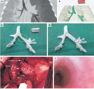
A two-month-old baby has saved his life upon being implanted with a splint in the trachea in a hospital in Michigan (USA), which was reproduced in a 3D printer, as published today in The New England Journal of Medicine.

The newborn suffered constant attacks caused by a syndrom called tracheobronchomalacia, which prevents oxygen from reaching the lungs due to a problem in the trachea problem, as diagnosed by doctors who have published the study. In view of the situation, doctors decided to print a kind of splint, which reproduced the tracheal tube based on a tomographic image of the child's airway.

The 3D reproduction is made with a material called polycaprolactone, bioabsorbable by the human body in three years, so the patient doesn't need another surgery to have the implant removed. The implant was reproduced on the printer in less than a day.

According to doctors, when the implant is removed naturally, the child's lungs and airways will be developed enough to stay open by themselves. After inserting the tube, doctors kept the baby on life support for 21 days, after which the baby was allowed to leave the hospital. One year after implantation, there have been no rejection or any problem with breathing.

This case demonstrates, doctors conclude in the study, that the combination of high resolution images plus the computer design and biomaterials for 3D printing can facilitate the creation of precise anatomical implants. The study has been signed by Scott Hollister and Richard Ohye from the University of Michigan, and Marc Nelson from Akron Children's Hospital.


The three-dimensional printers are making headway in medicine, but for now the applications are very limited. Initially, they were used to design conventional solid materials intended to design personalised implant prosthesis. One of the benefits is the ability to create custom models at lower prices than conventional industrial processes. The next step, as the case published today in the New England Journal of Medicine, is to design objects with a biological material such as the biodegradable polymer employed in the intervention to reproduce the trachea fragment that the patient was missing.

The ability to create these structures combined with the manipulation of stem cells could lead in the future to the creation of artificial organs. At least, scientist are working in this direction. A biological mold would be the basis to provide a three-dimensional structure to the organ. The outer casing would be scattered on cells which would colonize the structure resulting in specialized cells of the organ concerned (cardiomyocytes in the case of the heart, hepatomiocitos for the liver...).

This possibility remains part of science fiction. What is beginning to be a reality is a new generation of 3D printers that generates human tissue. The company Organovo announced last April at the Experimental Biology Conference in Boston an equipment capable of producing tiny liver tissue samples (about 20 cell layers thick). This printer uses biological ink which in this case is made of three types of cells: hepatocytes, stellate cells and cells from the walls of blood vessels, obtained from waste coming from transplants and surgical interventions. Currently, the main use of these miniature livers is to test the effectiveness and impact of new drugs, but it may be the first step in a future technology able to print of organs for transplantation.

http://www.nejm.org/doi/full/10.1056/NEJMc1206319
Tuesday, 14 May 2013

The most advanced bioartificial heart


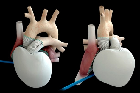
The French company Carmat has been authorized to begin implementing its new human artificial heart, considered one of the most advanced to date. Although only some pilot tests have been carried out so far, the new artificial organ will be used for the first time in four hospitals in Belgium, Poland, Slovenia and Saudi Arabia.

This model is considered the latest generation of artificial heart, and although there are many advantages that have been observed in the tests, it is also true that there is little experience with their use. Unlike mechanical artificial hearts, which consist of a pump that is connected to the heart or implanted in the chest to help or replace a failing heart (without removing it), bioartificial hearts are designed to completely replace the original organ.

Una imagen del nuevo dispositivo.| Carmat
New bioartificial heart (copyright Carmat)

The device has a price of 200,000 euros and a battery life of about 14 hours. Moreover is manufactured with a series of materials that reduce the risk of thrombi to the patient, resulting in less need for anticoagulant therapy. Although it is early to assess what the outcome will be, the Carmat heart presents already some advantages compared to its predecessors: it is a smaller model (it weights just one kg) and it comes with a SW that controls it efficiently (the artificial heart regulates itself very well and knows how much blood is necessary to pump at all times).

In a joint statement, cardiac surgeons of the four hospitals have declared that "terminal heart failure is a major public health problem that crosses borders. This artificial heart is a true innovation for the medical community. We are eager to sart early implementations and analyze its potential benefits for our patients". When talking about this artificial heart, we are probably talking about the future treatment of heart failure.

Thursday, 2 May 2013

A tattooed electronic circuit to monitor your body


The materials scientist, John Rogers, and the company MC10 BioStamp have developed a temporary tattoo with flexible electronic circuits that sticks directly to the skin to monitor the user's health. BioStamp patch is a fine mesh with electronic circuits which can control temperature, hydration and strain.

So far we only knew about the physical activity monitors, which have slowly been appearing on the market. BioStamp not only can monitor your activity, but control your vital signs and physical needs. The MC10 company team is now working on the integration of wireless power sources and communication systems to transmit the collected information to a Smartphone, so the BioStamp can also warn you about your physical and health needs. As an example, imagine your son of 11 months has a fever and it's time to sleep. You can use the BioStamp to have your SmartPhone alerting you if the fever rises overnight.


Rogers himself suggests that his "epidermal electronics" could be developed for use in healthcare, to monitor patients without tying them to large machines . Another possible application might be as a patch that allows an athlete to monitor their vital signs, blood pressure... as well as to know when and how much to hydrate for optimal performance.

BioStamp of MC10 is like a temporary tattoo, flexible and comfortable, formed by depositing gold electrodes and wires that are just a few hundred nanometers thick onto silicon wafers. Previous versions were made placing a patch elastomeric afterwards as a reinforcement, but the latest prototype is applied directly to the skin using a rubber stamp. It is applied with a spray coating which makes it durable and resistant to water enough to withstand the sweat or washing with soap and water. It lasts up to two weeks before the natural skin exfoliation.